Clearly, you're in good hands here.
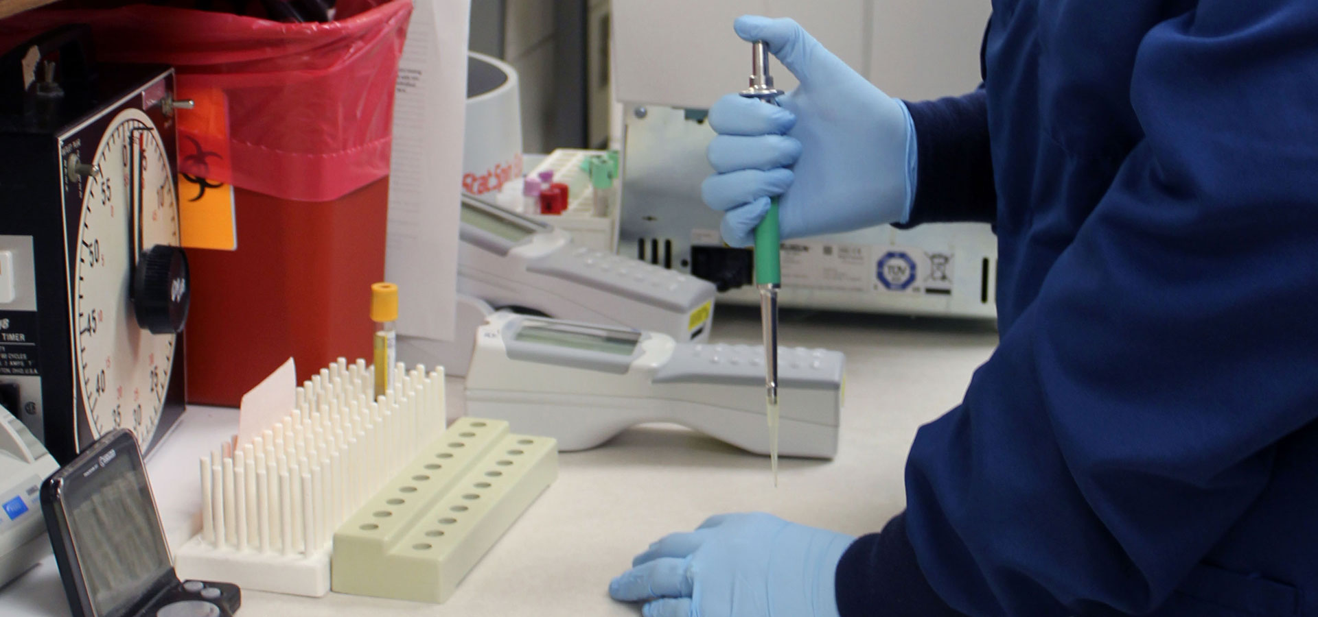
Our Radiology department is proud to offer a comprehensive selection of tests. We also partner with Diagnostic Radiology, P.C. to provide on-site care from experienced radiologists to our patients several days each week.
3D and Digital Mammography
We are proud to offer our patients state-of-the-art 3D mammograms. You can schedule your routine mammogram in your MyChart account, here online if you don't have a MyChart account, or by calling us directly at 712-243-7450. You can also take part in one of our quarterly Mammos & Mocktails evening events and make your mammogram more fun, with no extra cost. Learn more about Mammos and Mocktails and how to schedule your appointment here.
Bone Densitometry
Bone Densitometry, or DEXA (dual-energy x-ray absorptiometry), is the standard of care to measure bone loss and diagnose osteoporosis. The exam is quick, noninvasive and painless.
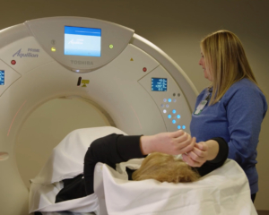
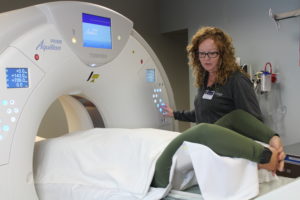
CT Scans
Cass Health has a state- of-the-art computerized tomography (CT) scanner with certified technicians available 24/7. CT scans provide a clear and specific image of internal organs, and can view a large portion of the body. The images are produced very quickly, which can be critical in emergency situations.
Digital Radiography
Digital radiography provides clear images, exposes patients to less radiation, and decreases patient wait time. Images are transmitted electronically, allowing healthcare providers to view them quickly.
Echocardiograms
An echocardiogram (echo) is a test that uses ultrasound to make pictures of your heart. Your doctor may use an echo test to look at your heart’s structure and check how well your heart functions. This test can help show your doctor:
- The size and shape of your heart, and the size, thickness and movement of your heart’s walls..
- The heart’s pumping strength.
- If the heart valves are working correctly.
- If there is a tumor or infectious growth around your heart valves.
Fluoroscopy
Fluoroscopy is an image of moving body structures, like an x-ray movie of the inside of your body. This allows your physician to see the internal structure and function, such as the movement of your throat as you swallow. Fluoroscopy images aid in both diagnosis and treatment therapies.
Interventional Radiology
Interventional Radiology uses medical imaging (CT, fluoroscopy, ultrasound) to assist the radiologist while performing minimally invasive procedures, such as biopsies. During a biopsy, a small sample of tissue is removed from the body and is sent on for further testing. Some of the more common biopsies that we perform include breast, thyroid, liver, lungs, and bone marrow. We also offer radiofrequency ablation to help reduce nerve pain.
MRI
MRI is a very precise method of imaging human anatomy. Through the use of a strong magnetic field, radio signals, and a computer, an educated technologist is able to construct anatomic images. MRI procedures do not involve radiation. MRI can be done on numerous body parts. Some of the most common scans are done to evaluate nervous system and musculoskeletal disorders. These disorders can include multiple sclerosis, tumors, stroke, injury, and diseases affecting tendons, ligaments, and cartilage.
Nuclear Medicine
Your healthcare provider may recommend a nuclear medicine procedure to diagnose or treat a health problem. Nuclear medicine is used to show how the organs or tissues are functioning. For most diagnostic procedures, a tracer, which contains the radioactive material, is injected, swallowed, or inhaled. Then the radiologist uses a radiation detector to see how much of the tracer is absorbed or how it reacts in the organ or tissue. This will give the provider information about how well it is functioning. Common uses of nuclear medicine for diagnosis include: scans of the heart, lung, kidneys, gallbladder, and thyroid.
PET Scans
PET scans use radioactive materials to show how tissues and organs are functioning and are typically used during cancer treatment. PET Scans are available on a mobile basis through Shared Medical Services.
Stereotactic biopsy
During a stereotactic biopsy, a special mammography machine uses x-rays to help guide the radiologist's instruments to the site of the abnormal growth. Stereotactic mammography pinpoints the exact location of the abnormality by using a computer and digital x-rays taken from two different angles. Using these computer coordinates, the radiologist inserts a needle through the skin, advances it into the lesion, and removes small tissue samples. This service is provided at Cass Health in partnership with a mobile unit from Health Enterprises.
Ultrasound
Ultrasound is a diagnostic procedure that uses high frequency sound waves to create images, and no radiation is involved in ultrasound imaging.